Schematic Drawing Of The Microscopic Structure Of Cardiac Mu
Cardiac muscle myocardiumleft ventricle Microscopic anatomy of cardiac muscle – ocr a level biology and chemistry Cardiac muscle cells gif microscopic mm ndsu pubweb edu
Solved 180 Chapter 11 The Cardiovascular System 4. Figure | Chegg.com
Cardiac muscle ventricle microscopic microscopy electron Human structure virtual microscopy Histology a464
Cardiac muscle – tutorial – histology atlas for anatomy and physiology
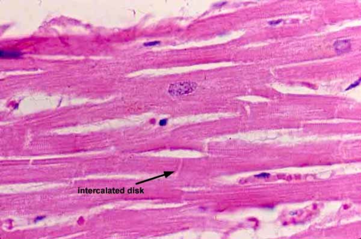
Cardiac muscle section skeletal stain tissue longitudinal human plate fluid phosphotungstic zenker acid hematoxylin anatomyatlasesCardiac muscle anatomy microscopic sarcolemma cells cell heart gap intercalated junctions muscles function discs tubules system tissue cardiovascular myocardium blood New page 5 [galeps.org]Cardiac vessels histology integument.
Histology lymphatic lymph valve vessels tissue group structure connective cardiac arteriole muscle identify weebly cardiovascular venule system smoothCardiac muscle tissue A464 muscle cardiac histology indiana medsci edu a560Muscle cardiac histology virtual structure human.

Cardiac muscle
Cardiac tissues annotatedCardiac muscle micrograph tissue lm regents provided michigan 1600 medical found university heart only school Plate 5.76: cardiac muscleCardiac muscle brief movement introduction support.
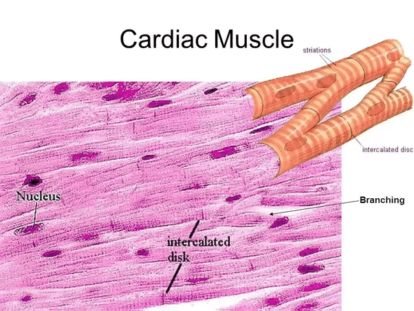
Microscopic structure schematic cardiovascular cardiacHuman anatomy: an overview of smooth, cardiac and skeletal muscles Muscle cardiac muscles human anatomy microscopic skeletal smooth cells appearance overviewOurlad's paradigm: muscle tissue.
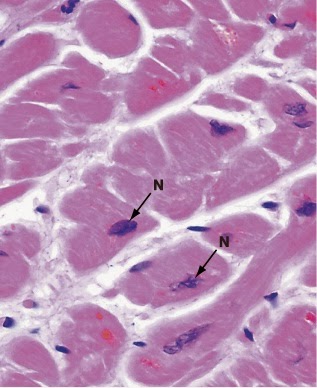
This image is a micrograph of cardiac muscle cells.
3. cardiac muscle tissueCardiac muscle micrograph cells Cardiac muscle – veterinary histologyGroup. 3: histology.
Solved 180 chapter 11 the cardiovascular system 4. figureCardiac cardiomyocytes intercalated disks histology nuclei veterinary pressbooks ohiostate centrally placed branching Muscle cardiac section cross tissue hpo ourlad paradigmMuscle cardiac microscopic longitudinal.

Cardiac microscopic reviewing fibers intercalated photomicrograph striations nucleus accompanying term 775x lm disc
Brief introduction on movement and support « simplebiologyMuscle cardiac muscles heart examples microscope under tissues function tissue cells nerve quora branching microscopy shape walls google form Cardiac muscle fig properties general microscopic structure ppt powerpoint presentationCardiac mm.
What are examples of cardiac muscles?Pre-lab activity 3: reviewing the microscopic stru... .


Cardiac muscle – Veterinary Histology
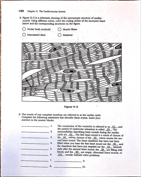
Solved 180 Chapter 11 The Cardiovascular System 4. Figure | Chegg.com
![New Page 5 [galeps.org]](https://i2.wp.com/galeps.org/jadams/biol 2212/images/Tissues/Annotated muscle/Cardiac muscle annotated.jpg)
New Page 5 [galeps.org]

Cardiac Muscle
Human anatomy: An overview of smooth, cardiac and skeletal muscles

This image is a micrograph of cardiac muscle cells.

PRE-LAB Activity 3: Reviewing The Microscopic Stru... | Chegg.com
What are examples of cardiac muscles? - Quora